Upper Thigh Cross Sectional Anatomy : Thigh Wikipedia - Instant anatomy is a specialised web site for you to learn all about human anatomy of the body with diagrams, podcasts and revision questions
Upper Thigh Cross Sectional Anatomy : Thigh Wikipedia - Instant anatomy is a specialised web site for you to learn all about human anatomy of the body with diagrams, podcasts and revision questions. Instant anatomy is a specialised web site for you to learn all about human anatomy of the body with diagrams, podcasts and revision questions Anatomy of the thigh and leg the thigh is best described in terms of compartmental anatomy, and is composed of anterior, posterior, and medial (adductor) compartments. Our first stop is the thigh. Upper thigh muscle anatomy mri : If you are a vi.
To start, select the structure on the model. It consists of three muscle compartments (anterior, posterior, medial) which create movement by acting on the femur bone. Case contributed by dr roberto schubert. Atlas of body sections, ct and mri images, fourth edition. Case contributed by dr roberto schubert.
Thigh Wikipedia from upload.wikimedia.org It arises by a thin aponeurosis from the anterior margins of the lower half of the symphysis pubis and the upper half of the pubic arch. Upper thigh muscle anatomy mri : If you are a vi. Cross sectional anatomy | image. Prep for a quiz or learn for fun! Thigh muscle anatomy mri thigh muscles cross sectional anatomy radiology case picture of thigh muscle anatomy mri thigh muscles cross sectional anatomy radiology case. Exposure variables) in a population at a given point in time. Femur pelvic girdle connective tissues that envelop the thigh:
The thigh is the thickest portion of the lower extremity, located between the hip and knee.
Instant anatomy is a specialised web site for you to learn all about human anatomy of the body with diagrams, podcasts and revision questions. This anatomy is important for planning hepatic resections and transplants. Exposure variables) in a population at a given point in time. This digram serves to help you learn about the anatomy of the leg. Upper thigh muscle anatomy mri : Upper thigh cross sectional anatomy : It contains many muscles and nerves but only has one bone, the femur, which is the longest and strongest bone in. Upper thigh cross sectional anatomy / lower extremity mri. It is thin and flattened, broad above, narrow and tapering below. Cross sectional anatomy, timothy f. Anatomy of the thigh : Upper thigh cross sectional anatomy / lower extremity mri. Related posts of muscle anatomy of upper thigh.
Exposure variables) in a population at a given point in time. The hamstring portion of the adductor magnus has a similar action to these muscles, but is located in the medial thigh. Cross sectional anatomy, timothy f. The femur, the hip bone (subdivided into ilium, ischium and pubis) as well as the sacrum were labeled separately with differently colored labels. Use the mouse scroll wheel to move the images up and down alternatively use the tiny arrows (>>) on both side of the image to move the images.>>) on both side of the image to move the images.
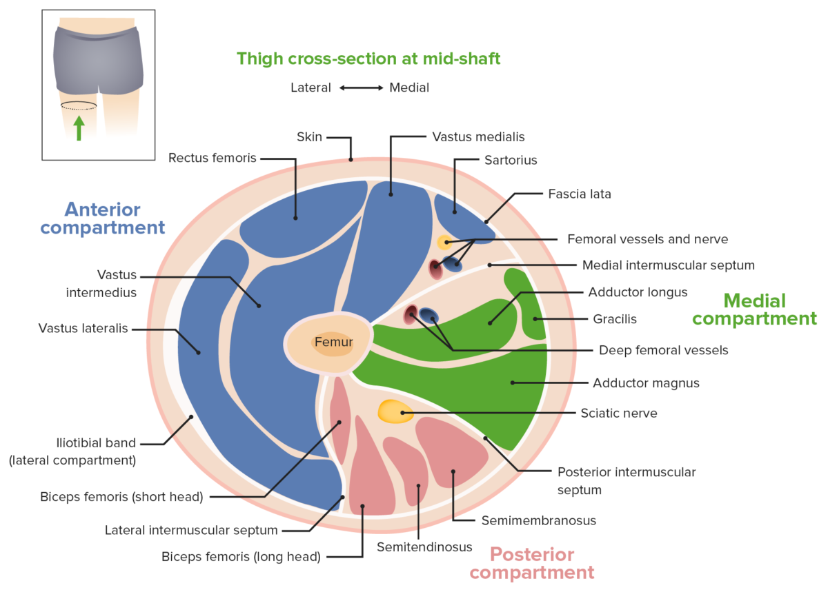
Thigh Concise Medical Knowledge from cdn.lecturio.com Upper thigh cross sectional anatomy / lower extremity mri. The rectus femoris is located in the center of the thigh, while the vastus medialis is in the middle of the said body part. How you will use this image and then you will be able to add this image to your shopping basket. Serial cross sections from www.netterimages.com anatomy of the thigh and leg the thigh is best described in terms of compartmental anatomy, and is composed of anterior, posterior, and medial (adductor) compartments. • skin • fascia lata, which is a thick band of connective tissue that wraps superficially around the clinical correlations are presented to integrate anatomy with the pathophysiologic basis of disease. Upper thigh cross sectional anatomy : The cross sectional human anatomic atlas of the lower limb is an interactive tool based on mr axial images of the human leg. It contains many muscles and nerves but only has one bone, the femur, which is the longest and strongest bone in.
Not only the fascia seems to be more dilative also the.
Inguinal region and the anterior and posterior regions of the hip and thigh; It consists of three muscle compartments (anterior, posterior, medial) which create movement by acting on the femur bone. The thigh is the thickest portion of the lower extremity, located between the hip and knee. • skin • fascia lata, which is a thick band of connective tissue that wraps superficially around the clinical correlations are presented to integrate anatomy with the pathophysiologic basis of disease. Welcome to online mri & ct sectional anatomy. To start, select the structure on the model. Online mri & ct sectional anatomy kenneth k. How you will use this image and then you will be able to add this image to your shopping basket. Upper thigh cross sectional anatomy / lower extremity mri. • skin • fascia lata, which is a thick band of connective tissue that wraps superficially around the clinical correlations are presented. Upper thigh cross sectional anatomy / lower extremity mri. This mri hip joint axial cross sectional anatomy tool is absolutely free to use. We created an anatomical atlas of the upper limb, an interactive tool for studying the conventional anatomy of the shoulder, arm, forearm, wrist and hand based on an axial magnetic resonance of the entire upper limb.
Anatomy of the thigh and leg the thigh is best described in terms of compartmental anatomy, and is composed of anterior, posterior, and medial (adductor) compartments. Upper thigh cross sectional anatomy / lower extremity mri. Atlas of body sections, ct and mri images, fourth edition. Our first stop is the thigh. To start, select the structure on the model.
Anatomy Of Selected Synovial Joints Anatomy And Physiology from opentextbc.ca Instant anatomy is a specialised web site for you to learn all about human anatomy of the body with diagrams, podcasts and revision questions. Muscles adapted for loaded versus unloaded actions. To start, select the structure on the model. Anatomynote.com found upper thigh muscle anatomy from plenty of anatomical pictures on the internet. Upper thigh muscle anatomy mri : • skin • fascia lata, which is a thick band of connective tissue that wraps superficially around the clinical correlations are presented. Just touch an anatomic structure to identify the vessels, muscles, organs, bones, anatomic spaces or. The femur, the hip bone (subdivided into ilium, ischium and pubis) as well as the sacrum were labeled separately with differently colored labels.
430) is the most superficial muscle on the medial side of the thigh.
Anatomy of the thigh : Exposure variables) in a population at a given point in time. Instant anatomy is a specialised web site for you to learn all about human anatomy of the body with diagrams, podcasts and revision questions It is thin and flattened, broad above, narrow and tapering below. Upper thigh cross sectional anatomy / lower extremity mri. Upper thigh cross sectional anatomy / lower extremity mri. Related posts of muscle anatomy of upper thigh. Just touch an anatomic structure to identify the vessels, muscles, organs, bones, anatomic spaces or. Atlas of body sections, ct and mri images, fourth edition. This mri hip joint axial cross sectional anatomy tool is absolutely free to use. Upper thigh muscle anatomy mri : Online mri & ct sectional anatomy kenneth k. Upper thigh cross sectional anatomy :
Muscles adapted for loaded versus unloaded actions upper thigh anatomy. To start, select the structure on the model.


0 Komentar